CryoStore diary - Sheyda Shapouri
Secondment at INRAE Rennes, France | October - December 2025After a series of drawn-out tearful goodbyes spread throughout Oslo and Ås, I set out for Rennes for my 2 and a half months stay at the Laboratoire de Physiologie et Génomique des Poissons at INRAE, where I would be learning cell culture techniques from the ground up. Coming from an academic background that distinctly lacked anything even resembling cell culture, I was a bit intimidated at the start, and the first day of sampling, preparation, and plating of our first culture was such a whirlwind of new information I barely managed to write down everything we were doing. As the weeks progressed though, I gradually went from watching things be done to doing some tasks under careful supervision, and before I knew it, I was managing the cells on my own!
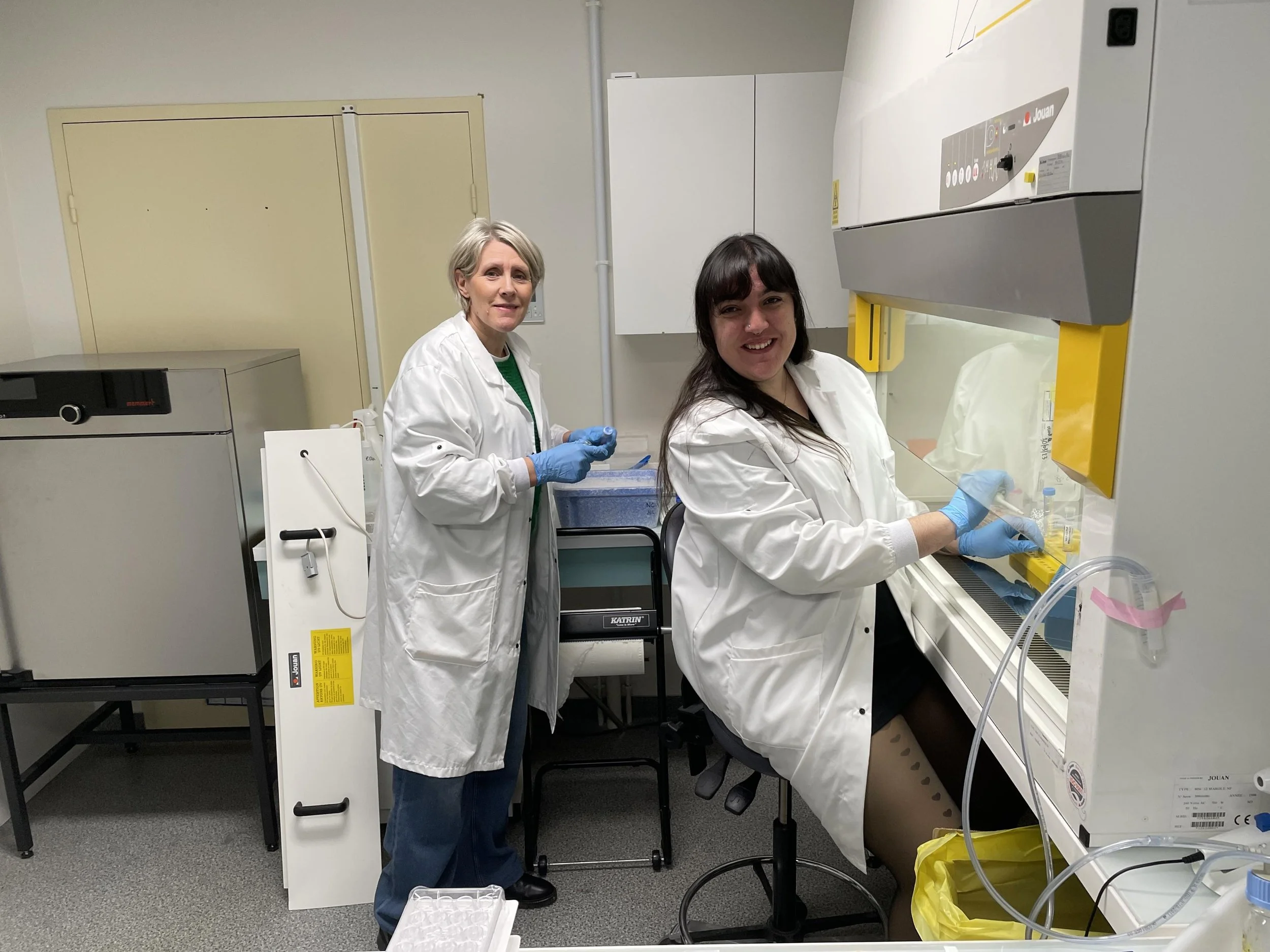
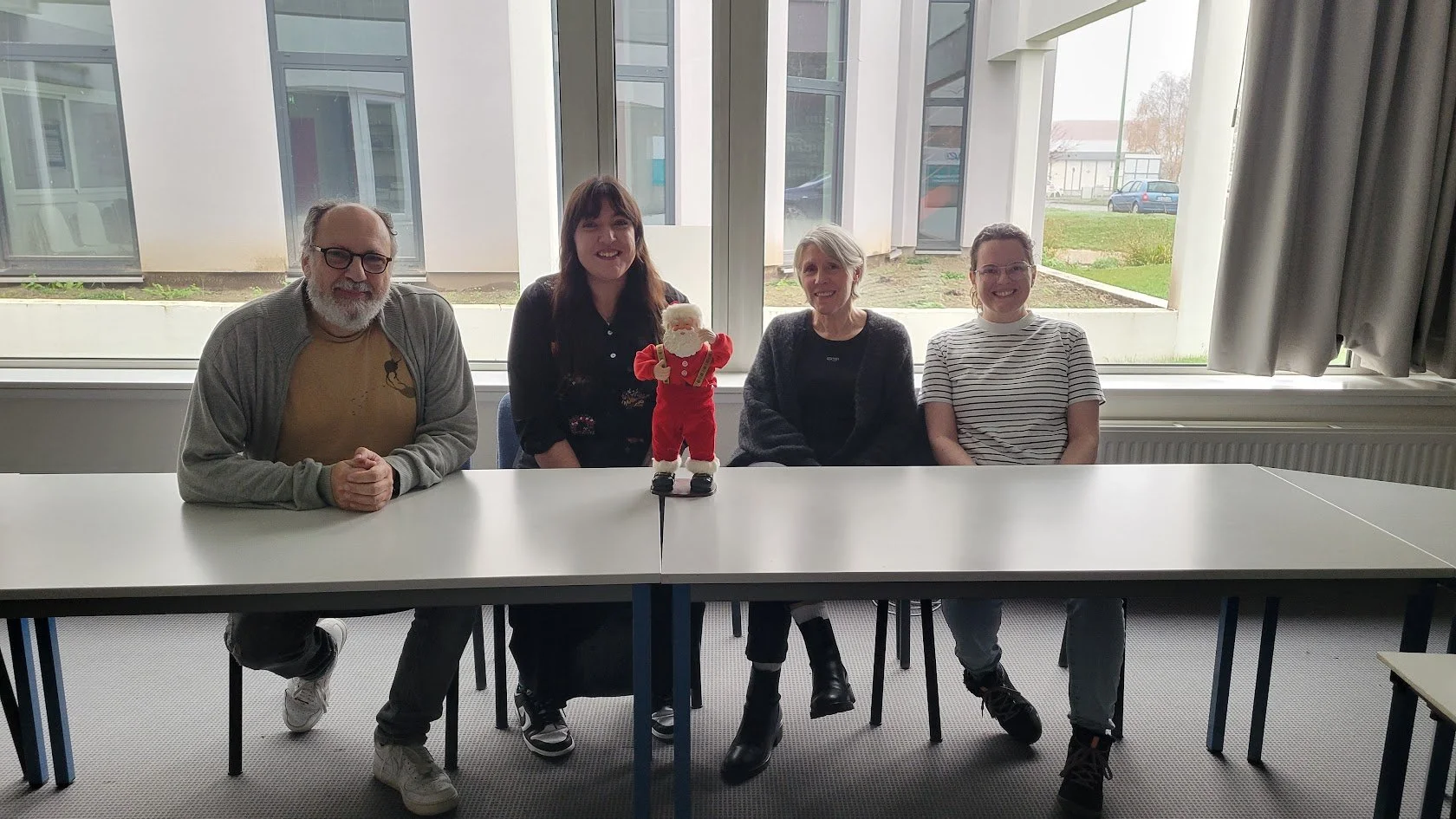
During the “close supervision” portion of my training, thank you Nathalie for all your help and patience!
On top of learning the ins and outs of cell culture, I also got to really savor the fruits of our labor and get a close look at the cells on the fluorescent microscope, a microscope I became incredibly familiar with throughout my time here. Because we used samples from transgenic zebrafish, the particular cells we were looking at fluoresced either red or green, depending on the cell type. This resulted in absolutely stunning images that I’ve been showing off to anybody who gives me a few minutes of their time, and material for PowerPoint slide backgrounds for the next six months at least!
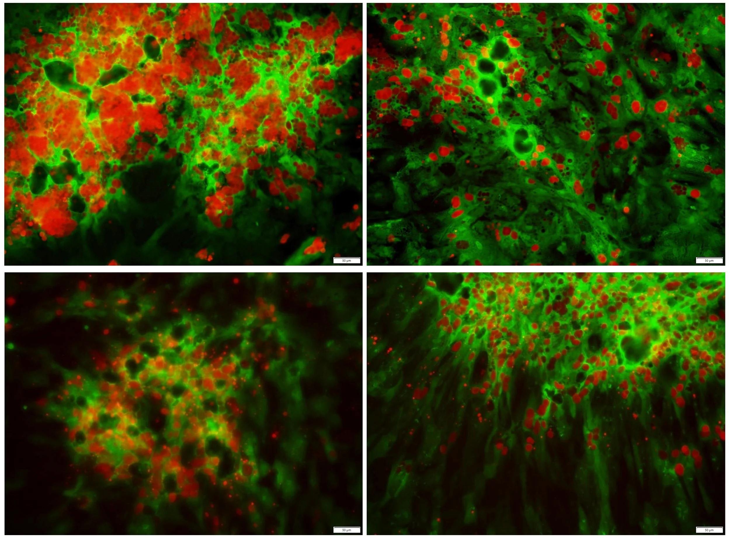
Photos from cultured zebrafish testicular cells. Sertoli cells (somatic cells in the testes) are in green and spermatogonia are in red.
I think I might be at the point where I’ve looked at green and red cells so much they’re showing up in my dreams, but I don’t even mind! I’ve taken it as a personal challenge to get a couple of really good artistic shots every time I have to go up and take pictures to track cell growth, and I’m really proud of the collection I’ve amassed so far. It was so fascinating to see the cells move and change as the culture progressed, and to be able to track their progress and overall status essentially in real time. I also got to learn about the behavior of the cells, the structures we were seeing them form into, and the functions of these structures right there at the microscope with my own images and cultures as a visual guide, which to me was much more valuable and easier to absorb than just reading papers and trying to retain everything.
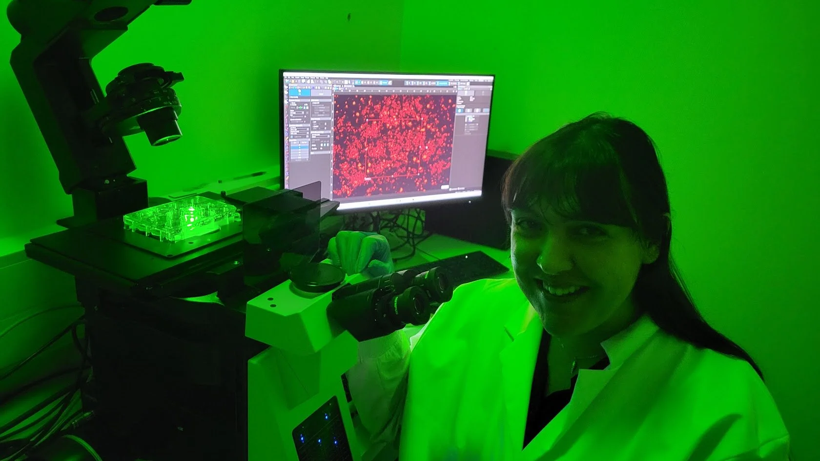
Me and my new best buddy, the fluorescent microscope.
I got to do a lot of observing and careful notetaking throughout the first culture we did, and when it came time for the second one it was on me to put all of the techniques I’d learned about in the first culture to the test. This culture was going to be entirely my responsibility, and in a way, a test to see if I was autonomous enough in the culture room to bring back everything I learned to NMBU and start fresh. Luckily for me, it all went well! We tested some new additives in the culture medium and took the cells through the full three weeks of culture with no contamination or other catastrophic events, a promising sign for my future cell culturing career.
In between caring for the cells, taking pictures, and preparing presentations and notes, I got to absorb a wealth of information about germ stem cells, their behavior, and their relationship to cells surrounding them both in vivo and in vitro. I almost couldn’t believe how much information I was absorbing every day, both technical knowledge I was directly putting into practice every time I tended to the cells and theoretical information about all the biological processes we were seeing unfold right in front of us. Also, regularly having to explain my progress in presentations to the group at LPGP and back at NMBU allowed me to really go through and examine everything I’d learned the week prior, making sure I was confident enough to explain it out loud and take questions, plus the added bonus of having a neat little timeline of my progress to be able to refer back to later.
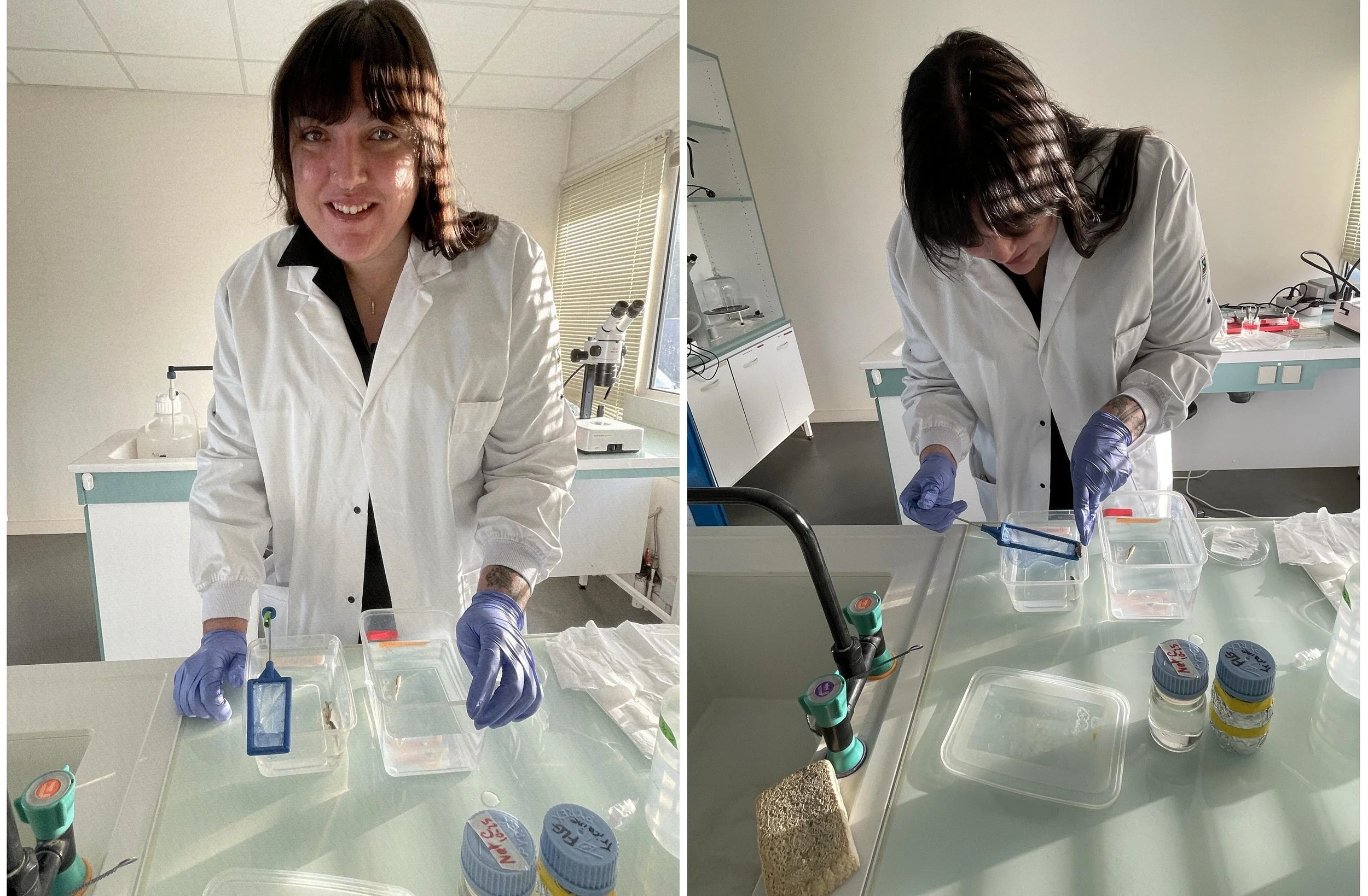
Preparing the fish for dissection on Day 1 of Culture 2
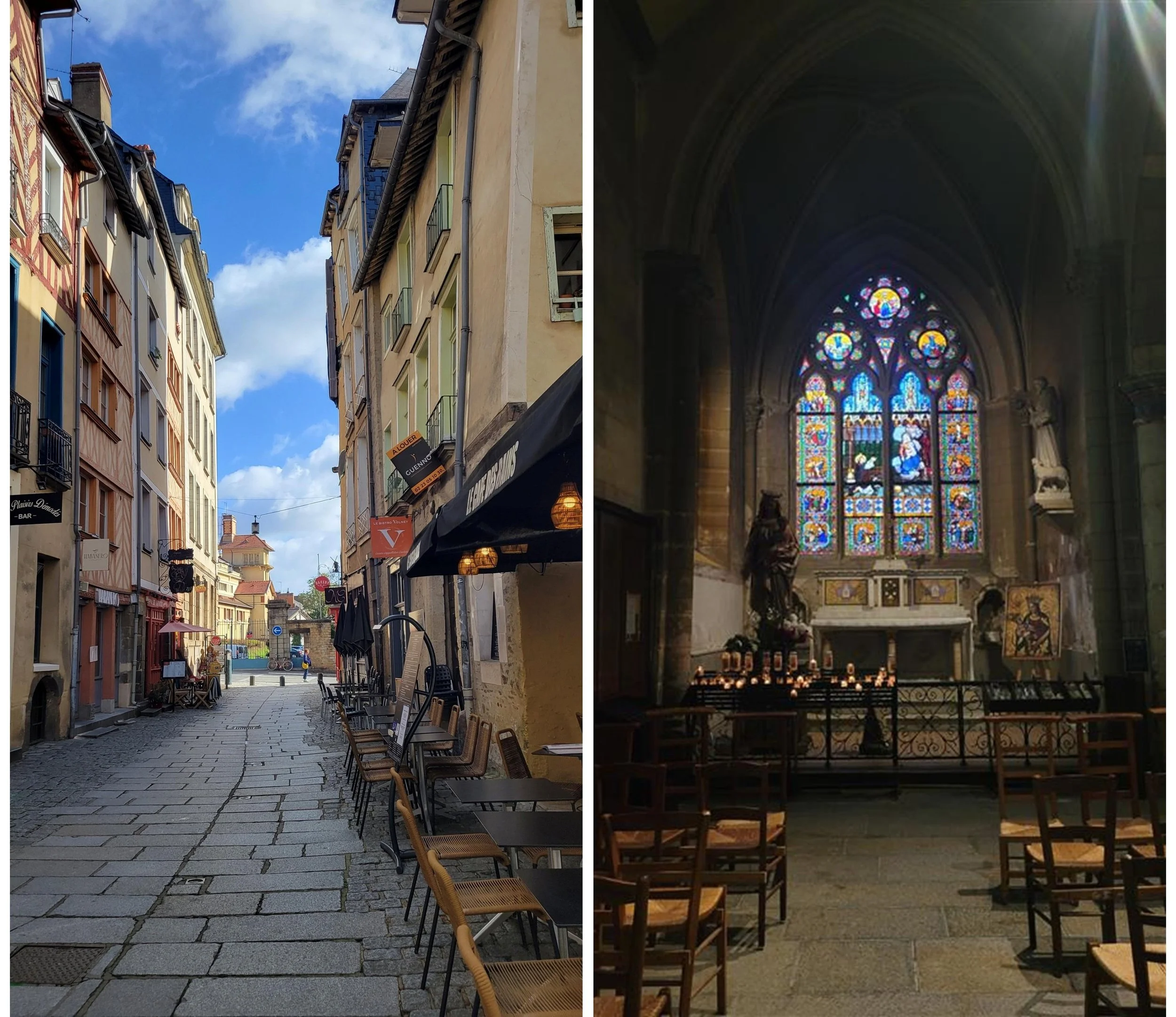
In my free time, I got to explore the twists and turns and charming old buildings of Rennes, mostly through my two main strategies for familiarizing myself with a new city: vintage shopping and local concerts! I’m pretty sure every cramped basement venue in town saw me at least once over the course of two and a half months, and it gave me a chance to meet people outside the institute and explore a side of Rennes I didn’t necessarily get to see during our summer school. Despite all the wet weather and usually coming home with soggy jeans, I made sure to wander the streets and weekend markets every chance I had (shoutout the cheese samples at Marché des Lices, I never bought any but I was always more than happy to try) and even put my best efforts into pretending to read the history of local cathedrals and historical buildings that were all in French.
Finally, I just want to thank everyone at LPGP again for turning this total newbie into a confident, full-fledged cell culture/fluorescent microscope/photography “expert” in such a short time. I learned so much more than I was even expecting to pick up, and I’m so excited to see where I can implement all these new techniques in my own research!
-Sheyda